
DIGESTIVE SURGERY
Gallbladder and Gallstones
Understanding Gallstones and Their Treatment
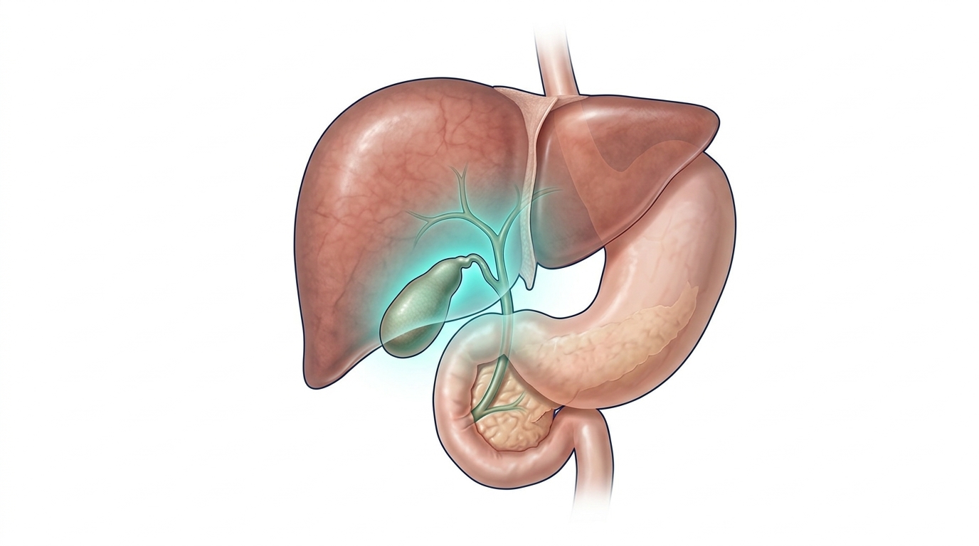
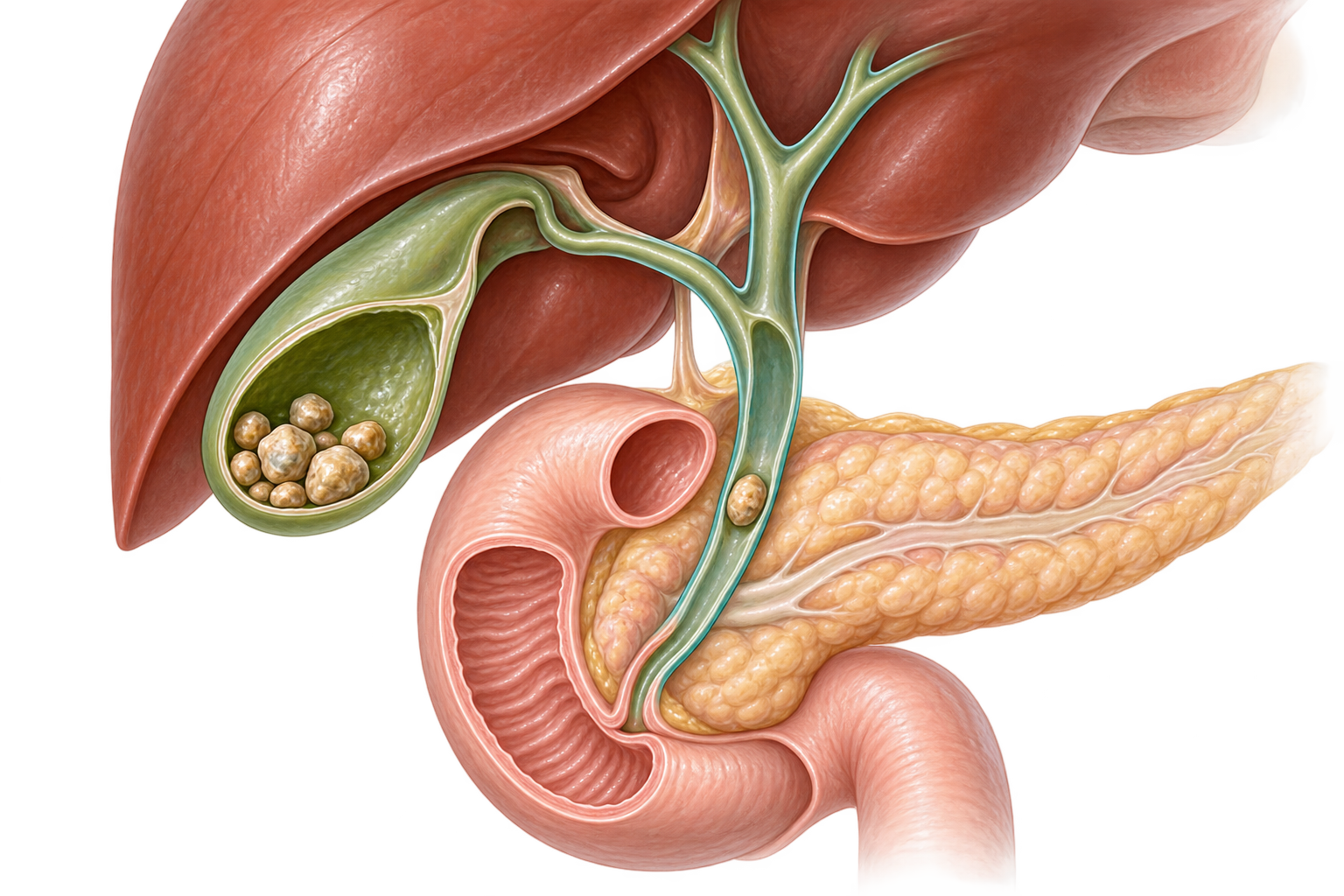
The gallbladder is a small organ located beneath the liver that stores bile.
Stones can form inside the gallbladder. They may remain completely silent or cause pain, inflammation of the gallbladder and, in some situations, complications affecting the bile ducts or the pancreas.
When gallstones become symptomatic or lead to certain complications, treatment may require removal of the gallbladder, known as cholecystectomy.
What Is the Gallbladder For?
A Reservoir for Bile
Bile is a fluid continuously produced by the liver that helps digest fats, among other functions.
The gallbladder is a small reservoir located beneath the liver. It stores and concentrates part of the bile between meals.
During digestion, the gallbladder contracts and releases bile into the bile ducts. The bile then reaches the duodenum, the first part of the small intestine.
It is important to understand that the gallbladder does not produce bile: the liver does.
The gallbladder is useful, but it is not essential. After it is removed, the liver continues to produce bile.
How Do Gallstones Form?
Stones That Develop Inside the Gallbladder
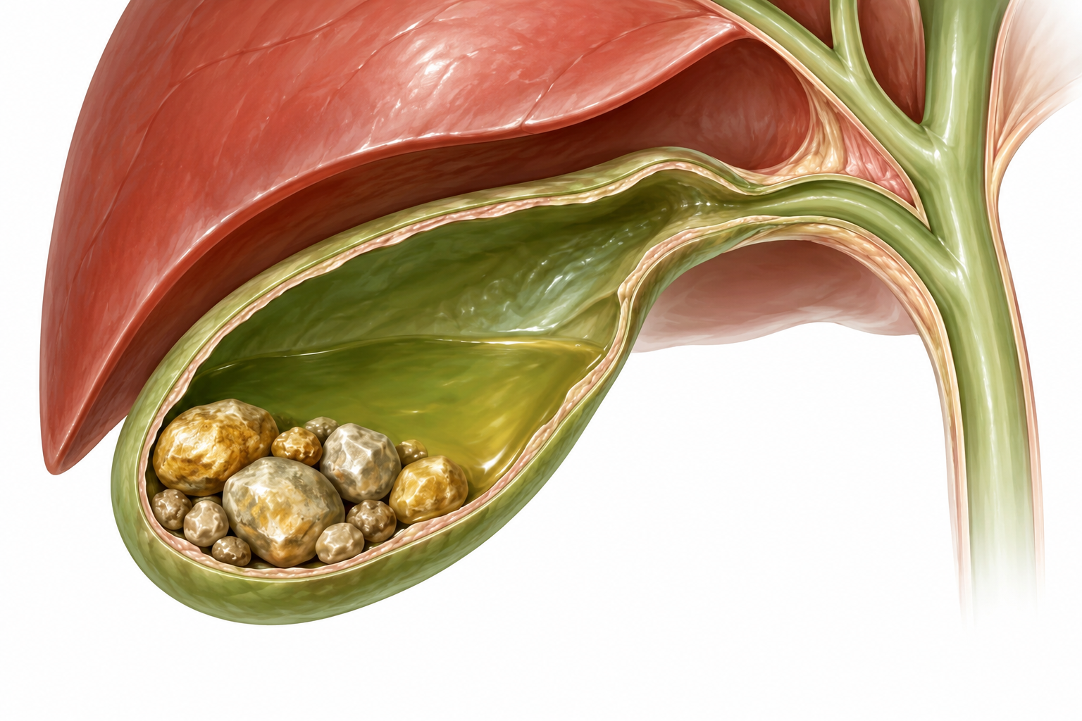
Gallstones, also known as biliary lithiasis or cholelithiasis, form when certain components of bile crystallize and aggregate inside the gallbladder.
Their number and size vary widely. A gallbladder may contain a single stone or several stones.
The presence of stones does not necessarily cause symptoms. Many are discovered incidentally during an ultrasound or another examination performed for a different reason.
The presence of stones in the gallbladder therefore does not automatically mean that surgery is necessary.
What Symptoms Can Gallstones Cause?
Biliary Colic Is the Most Characteristic Symptom
When a stone temporarily blocks the outlet of the gallbladder, it can cause pain known as biliary colic.
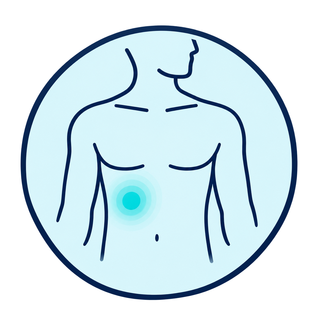
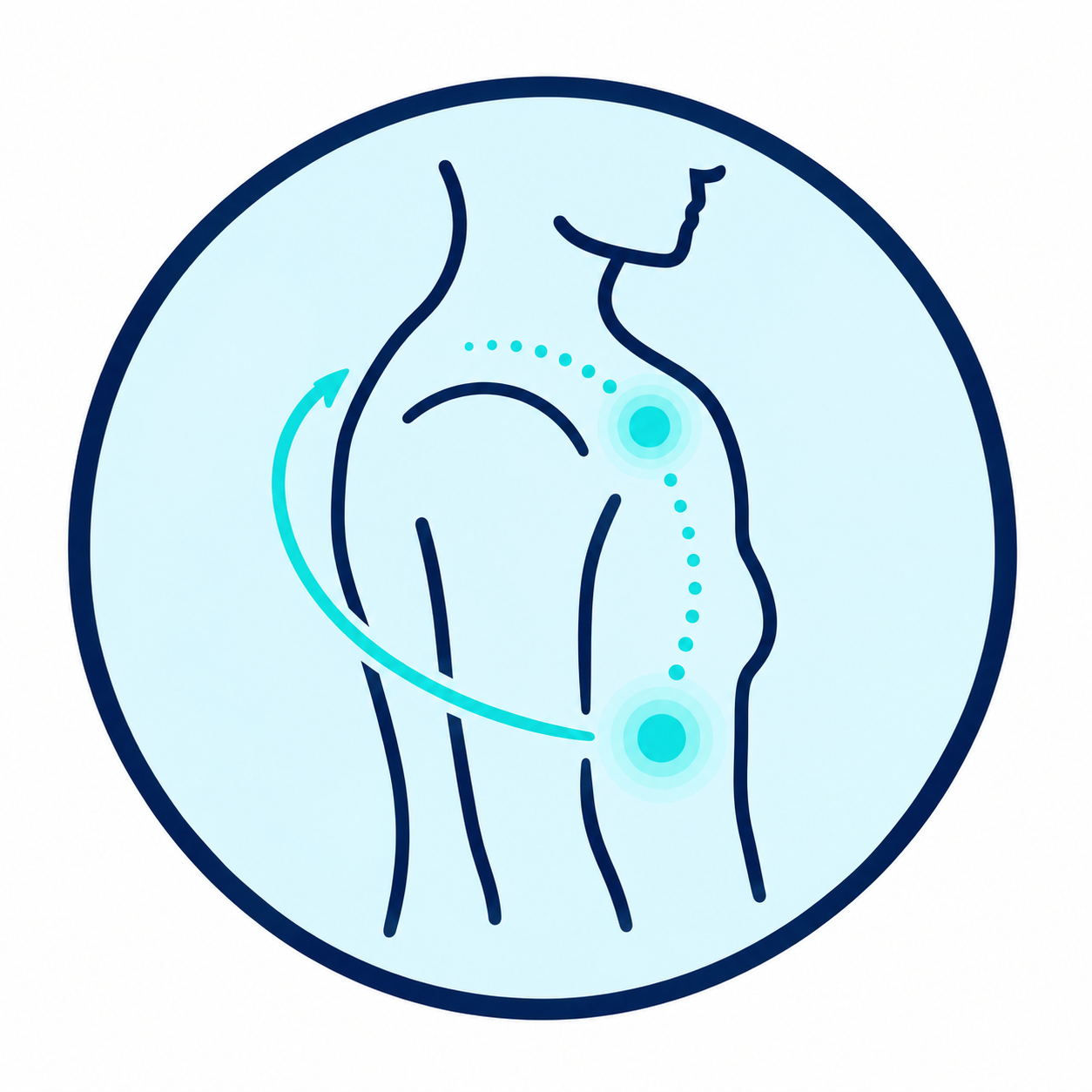
The pain usually appears in the upper right part of the abdomen or in the epigastric region. It may sometimes radiate to the back or the right shoulder. Episodes often occur after a meal and may be accompanied by nausea or vomiting.
On the other hand, some very common digestive symptoms — bloating, difficult digestion or diffuse abdominal discomfort — are not necessarily caused by gallstones. The presence of stones on ultrasound is not enough to prove that they are responsible for all digestive symptoms.
Abdominal Pain
Pain in the right upper quadrant or upper abdomen.
Radiation
The pain may sometimes extend to the back or the right shoulder.

Nausea
Nausea or vomiting may accompany an episode.

Painful Episodes
Episodes can appear suddenly and last from several dozen minutes to several hours.
What Complications Can Occur?
A Stone Can Sometimes Leave the Gallbladder or Cause Inflammation
Acute Cholecystitis
When a stone remains stuck at the outlet of the gallbladder, acute inflammation can develop. The pain is usually more prolonged and may be accompanied by fever.
Common Bile Duct Stone
A stone can leave the gallbladder and migrate into the common bile duct, called the choledochus. This is known as choledocholithiasis.
Cholangitis
Obstruction of the bile duct can, in some situations, be complicated by an infection of the bile ducts.
Biliary Pancreatitis
A small stone migrating toward the end of the bile duct can also contribute to triggering acute pancreatitis.
Significant and persistent pain, especially when accompanied by fever or jaundice, requires prompt medical evaluation.
How Is the Diagnosis Made?
Ultrasound Is Usually the First-Line Examination
The diagnosis is first based on symptoms and clinical examination.

Abdominal Ultrasound
Used in particular to look for stones in the gallbladder and signs of inflammation.
Blood Test
Used in particular to look for signs of inflammation or abnormal liver function tests.
Further Tests
When a common bile duct stone is suspected, further tests may be needed. Depending on the situation, this may involve MRI cholangiography or endoscopic ultrasound.
The work-up depends on the symptoms and the suspicion of an associated complication.
When Should Surgery Be Considered?
An Incidentally Discovered, Symptom-Free Stone Is Usually Not Operated On
The presence of stones in the gallbladder does not systematically lead to surgery.
Completely asymptomatic stones are generally monitored rather than operated on, except in specific situations. However, a cholecystectomy may be offered when the stones cause characteristic symptoms or certain complications.
It may notably be discussed in cases of: repeated biliary colic; cholecystitis; migration of a stone into the common bile duct; gallstone-related pancreatitis; or other specific situations.
The goal is to treat the gallbladder disease and prevent further stone-related episodes.
What Is a Cholecystectomy?
Surgical Removal of the Gallbladder
A cholecystectomy involves removing the gallbladder.
The procedure therefore does not simply consist of removing the stones.
Indeed, leaving the gallbladder in place after removing only the stones would expose the patient to the formation of new stones and the recurrence of symptoms.
When surgery is indicated, laparoscopic cholecystectomy is the usual approach.
How Is the Procedure Performed?
The surgery is performed under general anesthesia.
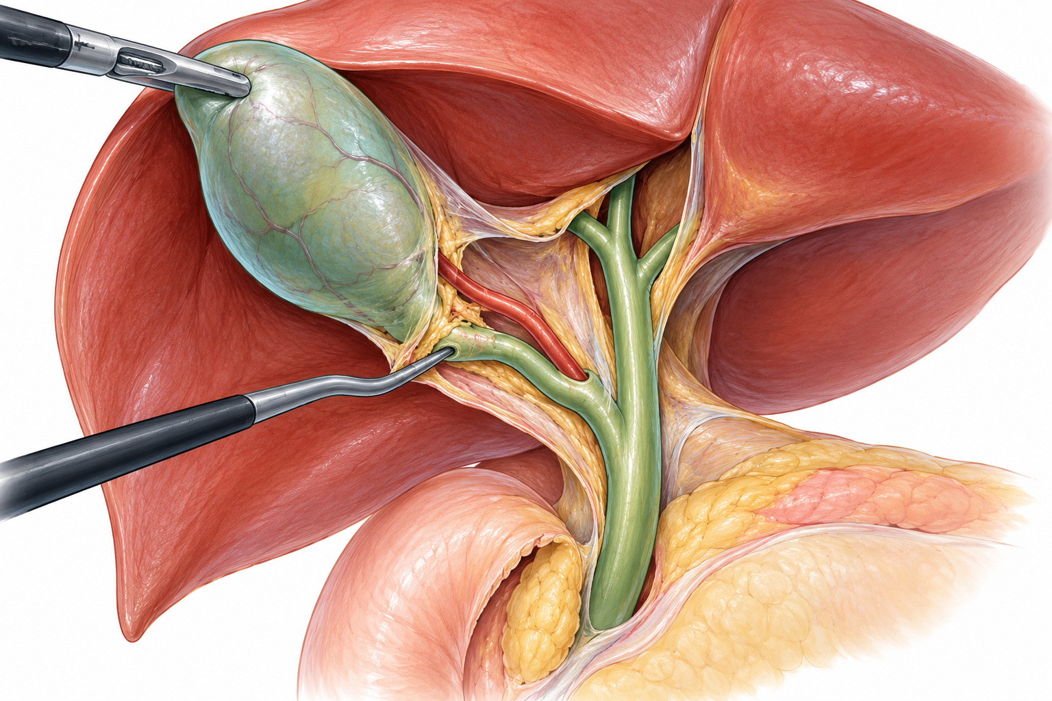
Several small incisions are used to introduce a camera and the surgical instruments.
The surgeon carefully identifies the anatomy of the gallbladder, the cystic duct and the cystic artery before controlling them.
The gallbladder is then separated from the liver and removed.
The safety of the procedure relies in particular on precise identification of the anatomy before dividing any structures; the “critical view of safety” is a reference strategy for this identification.
After the procedure, the liver continues to produce bile, which flows directly into the intestine through the bile ducts.
What Is Intraoperative Cholangiography?
Visualizing the Bile Ducts During Surgery
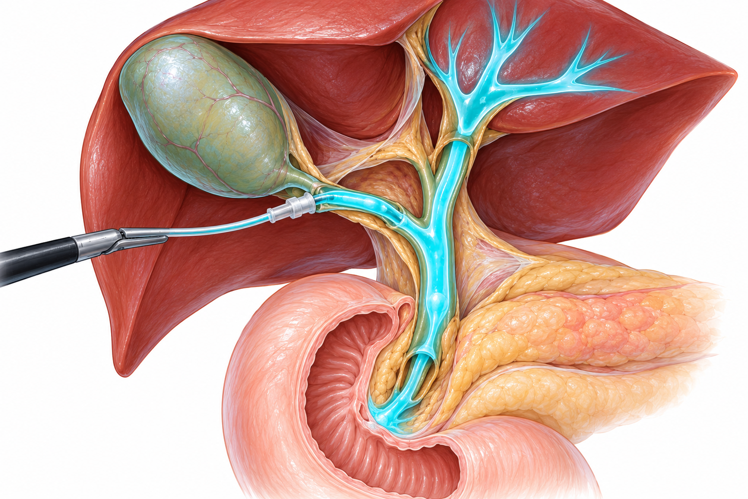
Intraoperative cholangiography is an examination performed during cholecystectomy.
A small catheter is introduced into the cystic duct and a contrast agent is injected into the bile ducts.
X-ray images then make it possible to visualize the anatomy of the bile ducts and the passage of contrast toward the duodenum.
Cholangiography can also help detect a stone present in the common bile duct.
Recent SAGES guidelines suggest the use of intraoperative cholangiography during cholecystectomies for benign biliary disease, but this is a conditional recommendation based on low-certainty evidence. Its value is particularly recognized when the anatomy is uncertain or a bile duct injury is suspected.
Cholangiography does not remove the gallbladder: it provides the surgeon with an image of the bile ducts during the procedure.
Why Look for a Stone in the Common Bile Duct?
A Stone May Have Left the Gallbladder Before Surgery
A stone initially formed in the gallbladder can migrate into the common bile duct.
It can sometimes be suspected before surgery based on symptoms, blood tests or imaging studies. In other situations, it may be identified during intraoperative cholangiography.
When a common bile duct stone is found, several treatment strategies are possible, either during or around the time of cholecystectomy. The choice depends in particular on the anatomical situation, the stone itself, the expertise available and how care is organized. Guidelines do not show a single strategy to be superior in every situation.
The gallbladder and the common bile duct are two different structures: removing the gallbladder does not automatically mean that a stone located in the common bile duct is removed.
Laparoscopy or Robot-Assisted Surgery
Two Possible Minimally Invasive Approaches
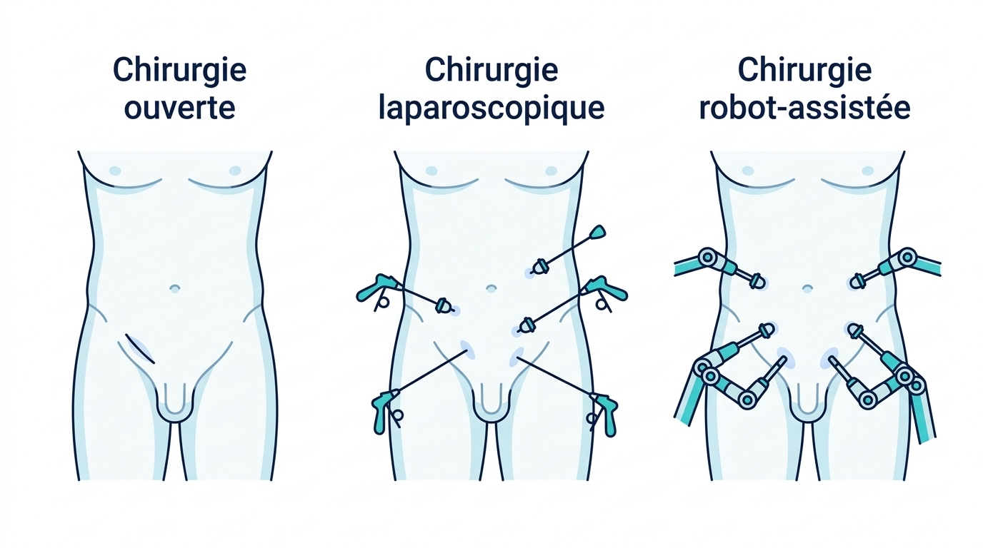
Cholecystectomy is usually performed using a minimally invasive approach.
Laparoscopy
The surgeon uses a camera and instruments introduced through several small abdominal incisions.
Robot-Assisted Surgery
Cholecystectomy can also be performed with robotic assistance in certain situations. The surgeon controls the instruments from a console. The robot does not perform any movement autonomously.
What Are the Risks of Cholecystectomy?
A Common Procedure That Remains a Surgical Operation
As with any surgical procedure, it is important to be aware of the risks associated with cholecystectomy.
As with any procedure, bleeding or infection can occur.
A bile leak can rarely occur after the procedure and may require further tests or additional treatment.
Injury to the common bile duct is a rare but important complication of cholecystectomy. Precise identification of the anatomy is therefore a central element of surgical safety.
A stone can sometimes remain, or be discovered later, in the common bile duct.
In some situations, continuing the procedure by laparoscopy may not be safe enough. Conversion to open surgery may then be necessary.
The priority is always the safety of the procedure. A change in the surgical strategy may be decided when the anatomy or local conditions require it.
After the Procedure
A Generally Gradual and Rapid Recovery
Getting up and moving is generally encouraged early after a cholecystectomy.
Eating can be resumed gradually, depending on comfort and the instructions given after the procedure.
It is generally not necessary to follow a special long-term diet simply because the gallbladder has been removed.
Daily activities are resumed gradually. Returning to work, driving, sport and physical effort depends on recovery and the type of activity.
Personalized instructions are given after the procedure, based on your situation and how the operation went.

Can You Live Normally Without a Gallbladder?
Yes, the Liver Continues to Produce Bile
After a cholecystectomy, the liver continues to produce bile.
The difference is that it is no longer stored in the gallbladder between meals: it flows directly from the liver to the intestine through the bile ducts.
The vast majority of patients can therefore return to their usual diet after the recovery period.
Some people may nevertheless experience temporary digestive changes after the procedure.
Removing the gallbladder therefore does not eliminate the liver, bile production, or the ability to digest normally.
A Personalized Approach to Care
Determining Whether the Stones Truly Explain the Symptoms
The discovery of gallstones does not automatically mean that surgery is necessary.
The consultation makes it possible to analyze the symptoms, the ultrasound findings and any associated complications.
When surgery is indicated, the strategy is tailored to the situation and to the possibility of associated involvement of the bile ducts.
The goal is to offer the necessary treatment without systematically attributing digestive symptoms to the gallbladder when they could have another origin.
Dr Pierre Fournier
General and Visceral Surgery
[Real photo of Dr Fournier — to be added once available]
Frequently Asked Questions:
No. Asymptomatic stones generally do not require a cholecystectomy, except in specific situations. Surgery is mainly considered when the stones become symptomatic or lead to certain complications.
No. The usual surgical treatment consists of removing the gallbladder along with the stones it contains, rather than simply extracting the stones from the gallbladder.
Yes. The liver continues to produce bile after cholecystectomy.
It is the removal of the gallbladder using a camera and instruments introduced through small abdominal incisions.
Intraoperative cholangiography makes it possible to visualize the bile ducts during the procedure through the injection of a contrast agent. It can, in particular, clarify the anatomy and reveal a stone in the common bile duct.
Practices can vary. Recent SAGES guidelines suggest its use during cholecystectomies for benign biliary disease, including a routine rather than selective strategy, but these recommendations are conditional and based on low-certainty evidence. They also specify that the decision should take into account the individual context, the surgeon’s experience and the resources available.
Several options exist for treating a common bile duct stone, either endoscopically or surgically depending on the situation. The strategy is individualized.
Since the gallbladder has been removed, stones can no longer form inside it. However, stones can rarely be present or form in the bile ducts.
A permanent specific diet is generally not necessary after a cholecystectomy. Eating is resumed gradually according to individual tolerance.
Yes, a robot-assisted approach is possible in certain situations. The robot remains entirely controlled by the surgeon.
Do You Have Gallstones?
The presence of gallstones does not systematically mean that surgery is necessary. A consultation makes it possible to determine whether the stones explain the symptoms, to look for a possible complication and to define the appropriate care.