
COLORECTAL SURGERY
Rectal Cancer
A personalized treatment based on tumor location and stage
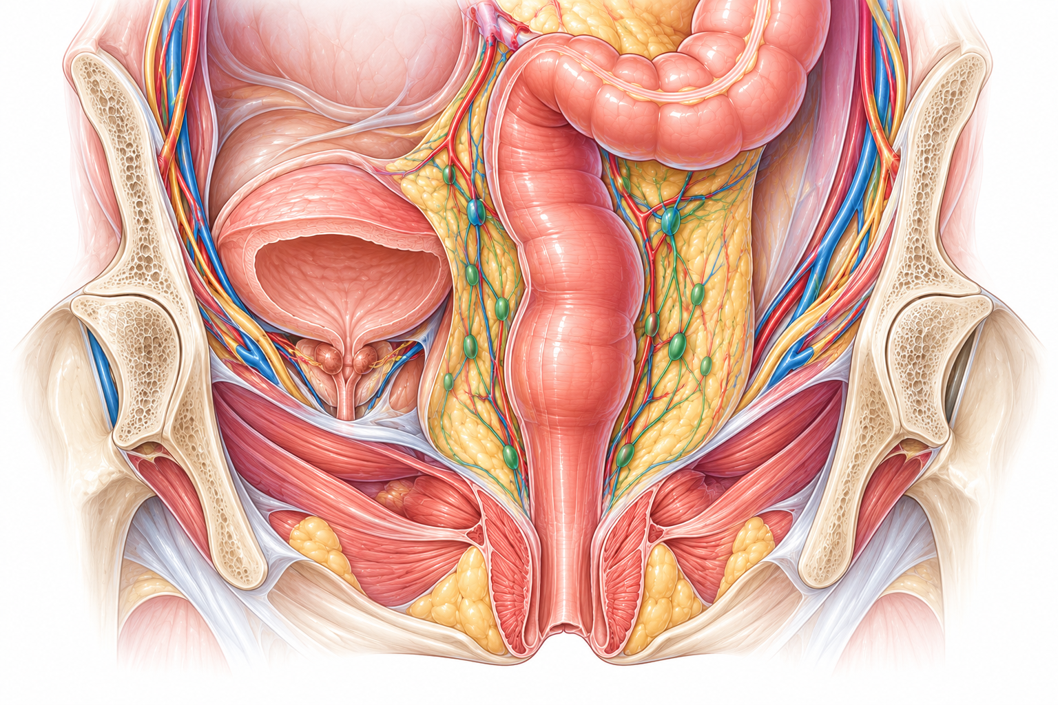
Rectal cancer develops in the terminal portion of the large intestine, located in the pelvis, just before the anal canal.
Its management differs significantly from colon cancer because of the rectum’s location within the pelvis and its proximity to the anal sphincter and neighboring organs.
The work-up makes it possible to precisely determine the tumor’s location, its extension, and its relationship with surrounding structures.
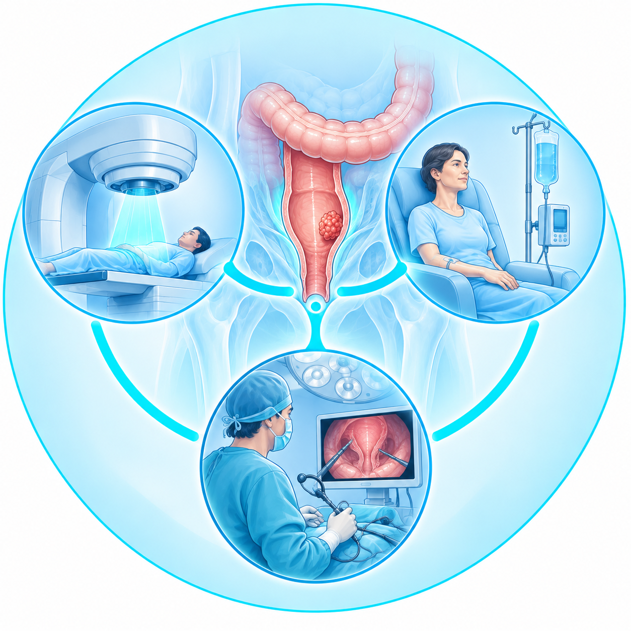
Depending on the situation, treatment may combine surgery, radiotherapy, and drug therapy in an order tailored to each patient.
What is the rectum?
The terminal portion of the large intestine
The rectum extends from the sigmoid colon and ends at the anal canal.
Unlike most of the colon, the rectum is located deep within the pelvis.
It lies close to several important structures, including:
- the bladder and urinary tract
- the genital organs
- the pelvic floor muscles
- the anal canal and sphincters
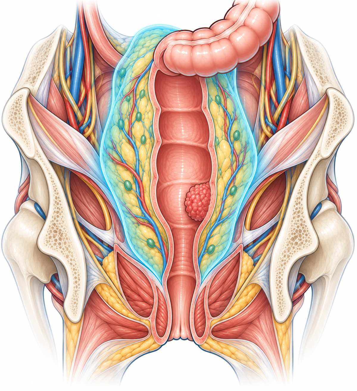
The rectum is also surrounded by fatty tissue containing blood vessels and lymph nodes: the mesorectum.
This anatomy explains why the work-up and surgery for rectal cancer have important specific features.
Colon cancer and rectal cancer: what is the difference?
Two nearby locations, but sometimes different strategies
Colon and rectal cancers both belong to the colorectal cancer family.
They nonetheless present important differences.
The rectum’s location within the pelvis means the tumor may lie close to:
- the anal sphincter
- the mesorectum
- the bladder
- the urinary tract
- the genital organs
- the pelvic floor
Local assessment must therefore be particularly precise.
For certain rectal tumors, treatment may also be given before surgery to optimize disease control.
→ Learn more about colon cancer
What are the symptoms of rectal cancer?
Sometimes subtle symptoms
Rectal cancer can be discovered during screening or a colonoscopy performed for another reason.
When present, symptoms may include:
- blood in the stool
- a persistent change in bowel habits
- frequent or unusual urges to have a bowel movement
- a feeling of incomplete emptying
- a change in stool shape
- fatigue or anemia
- sometimes pelvic discomfort or pain
These symptoms can have many other causes.
Blood in the stool or a persistent change in bowel habits nonetheless deserves medical evaluation.

How is rectal cancer diagnosed?
Colonoscopy and biopsies
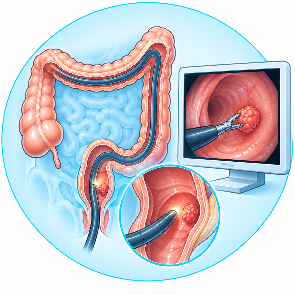
Colonoscopy allows examination of the colon and rectum.
When a rectal lesion is identified, biopsies are usually performed.
The samples are then analyzed under a microscope to determine the nature of the lesion.
This analysis confirms the diagnosis and clarifies certain characteristics of the tumor.
Examining the entire colon also makes it possible to look for any other lesions.
A clinical examination of the rectum can provide additional information, particularly when the tumor is low enough to be accessible.
Why perform a pelvic MRI?
An essential exam for precisely understanding the tumor
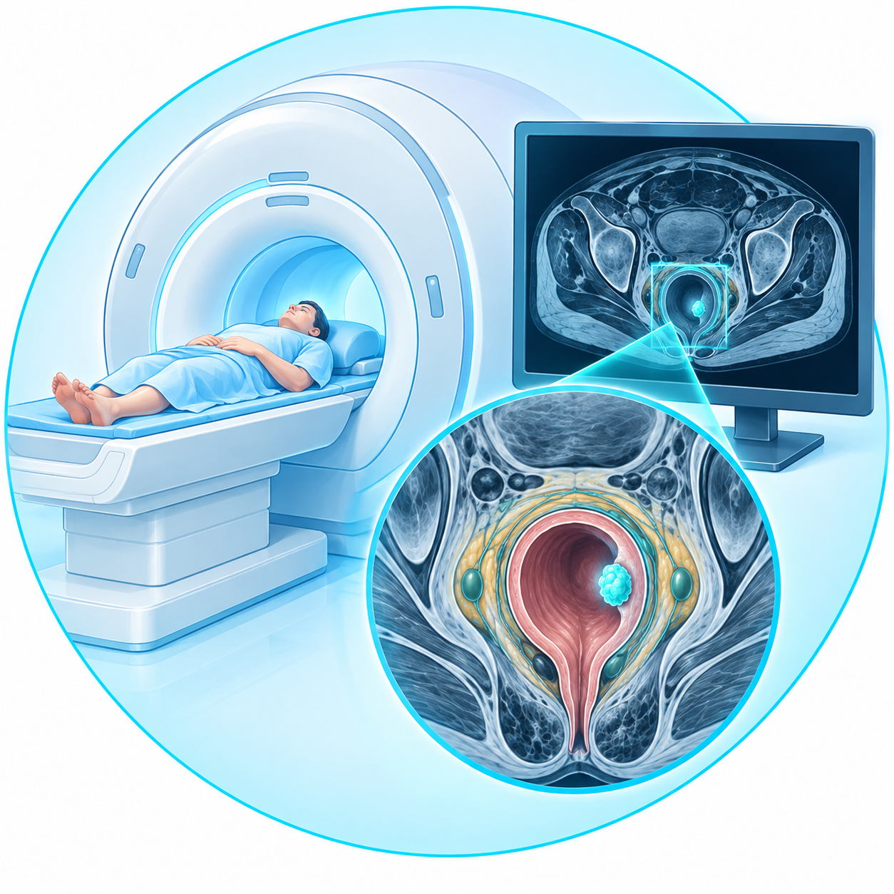
Pelvic MRI plays a particularly important role in the work-up of rectal cancer.
It allows precise study of the tumor and its relationship with surrounding structures.
It notably helps assess:
- the depth of tumor extension
- the mesorectum
- the regional lymph nodes
- the tumor’s proximity to the resection margins
- its relationship with the sphincter and pelvic structures when it is low
This information directly contributes to choosing the treatment strategy.
Staging work-up
Assessing the disease locally and at distance
The work-up does not stop at studying the rectum.
Chest, abdominal, and pelvic imaging also makes it possible to look for any distant spread of the disease.
Blood tests are performed and may include measuring CEA.
CEA alone cannot diagnose rectal cancer, but it can provide useful information for management and follow-up.
All the results together make it possible to define the stage of the disease.
How is treatment chosen?
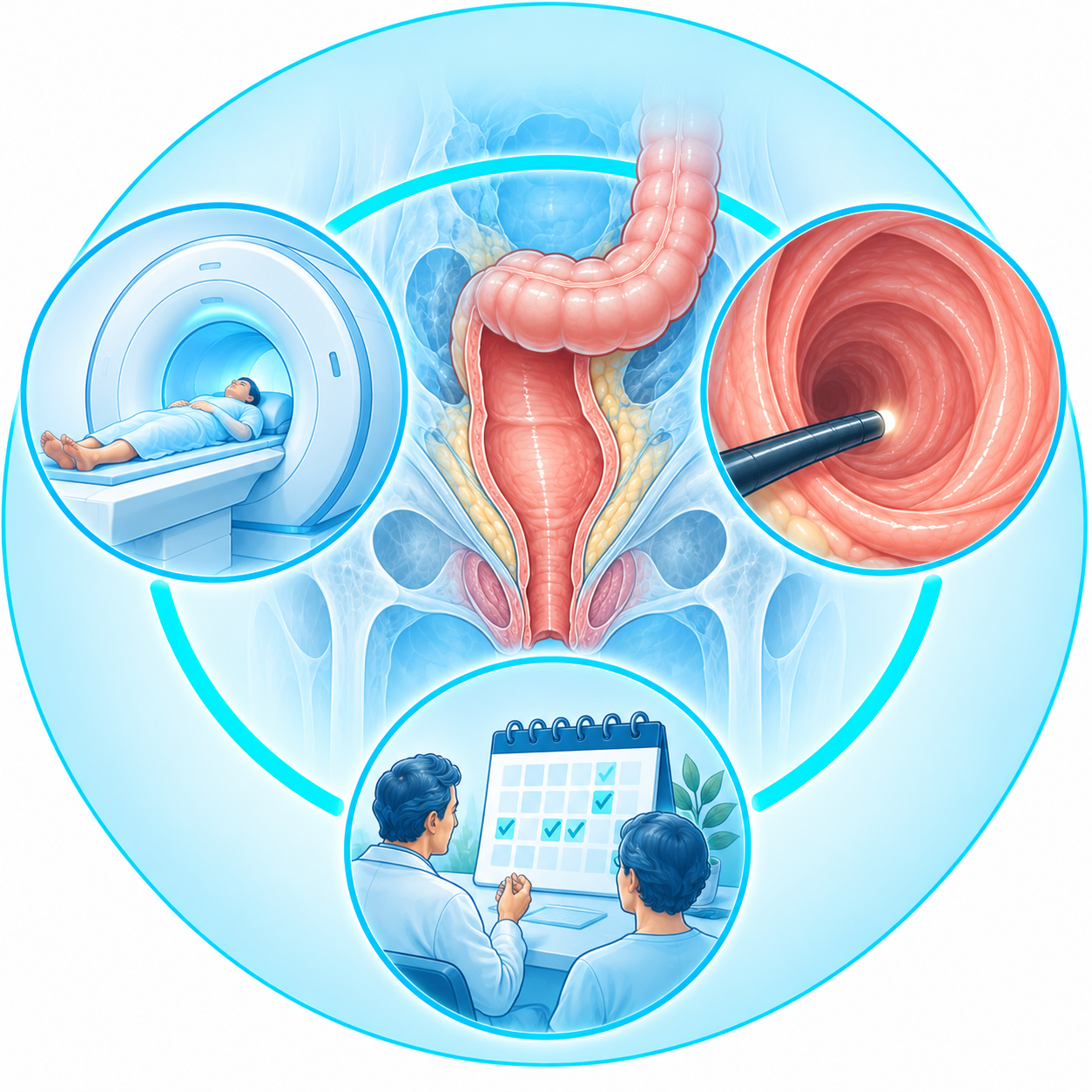
Not all rectal tumors follow the same pathway
Treatment depends on several factors:
- the tumor’s height within the rectum
- its depth of extension
- the lymph nodes
- its relationship with the mesorectum and neighboring structures
- any possible distant disease
- certain biological characteristics of the tumor
- the patient’s general health
Some early-stage tumors may be suitable for initial surgical treatment.
For other rectal cancers, treatment is given before surgery.
The strategy is therefore defined individually within a multidisciplinary framework.
Why sometimes treat before operating?
Reducing the risk of recurrence and optimizing the surgical strategy
For certain locally advanced rectal cancers, or those with specific features on MRI, preoperative treatment may be offered.
Depending on the situation, it may combine:
- radiotherapy
- chemotherapy
- chemoradiotherapy
- or a sequence combining several treatments before surgery
This is commonly referred to as neoadjuvant treatment.
In some situations, all planned chemotherapy and radiotherapy can be administered before surgery: this strategy is called total neoadjuvant therapy, or TNT.
The goal is not simply to “shrink the tumor.”
The strategy notably aims to improve local control and to treat any microscopic disease early.
What does surgery involve?
Removing the diseased rectum along with its lymphatic territory
When rectal resection is indicated, the goal is to remove the portion of the rectum containing the tumor with adequate oncologic margins.
For many mid- and low-rectal tumors, surgery also includes careful removal of the mesorectum surrounding the rectum.
This technique is called total mesorectal excision, or TME.
The mesorectum notably contains the blood vessels and lymph nodes associated with the rectum.
The quality of this dissection is an essential element of oncologic rectal surgery.
Can the sphincter be preserved?
Tumor location is decisive
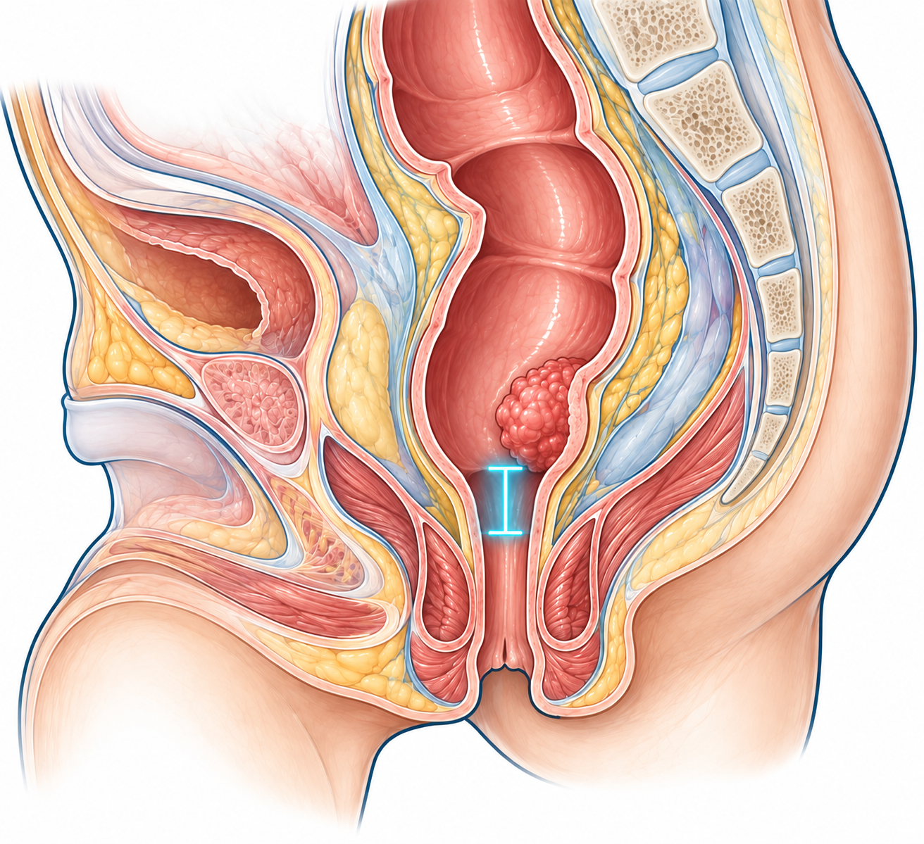
One of the key challenges of rectal surgery is determining whether the tumor can be removed while preserving the anal sphincter.
This possibility depends notably on:
- the tumor’s height
- its extension
- its relationship with the sphincter
- the response to any preoperative treatment
- the ability to achieve a satisfactory oncologic resection
- pre-existing sphincter function
When conditions are favorable, the diseased rectum can be removed and digestive continuity then restored.
In certain very low tumors, sphincter-preserving surgery is not oncologically or functionally appropriate.
How is digestive continuity restored?
Colorectal or coloanal anastomosis
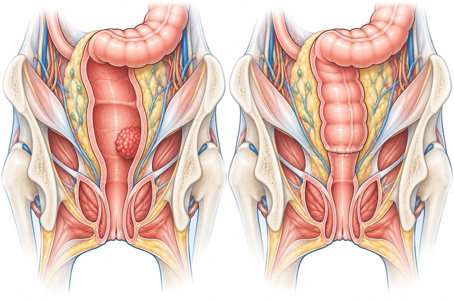
When the sphincter can be preserved, the remaining colon is connected to the remaining rectum or, for very low resections, to the anal canal.
This junction is called an anastomosis.
It can therefore be:
- colorectal
- or coloanal, depending on the level of resection
The goal is to create a well-vascularized digestive continuity without excessive tension.
Is a stoma always necessary?
No, but it can serve several purposes
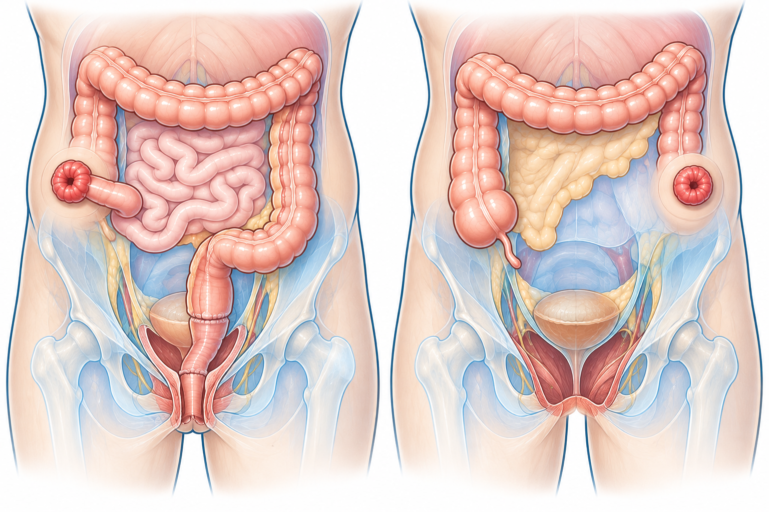
Whether a stoma is needed after rectal surgery depends on the type of procedure.
When a very low anastomosis is performed, a temporary diverting stoma may be offered in certain situations.
Its purpose is to temporarily divert the bowel while the anastomosis heals.
It can then be closed once the necessary conditions are met.
In other situations, particularly when the tumor requires removal of the rectum and anal sphincter, a permanent colostomy may be necessary.
It is therefore important to distinguish between:
temporary diverting stoma ≠ permanent stoma.
The strategy is discussed individually before surgery whenever possible.
Laparoscopy and Robot-Assisted Surgery
Minimally invasive approaches suited to certain situations
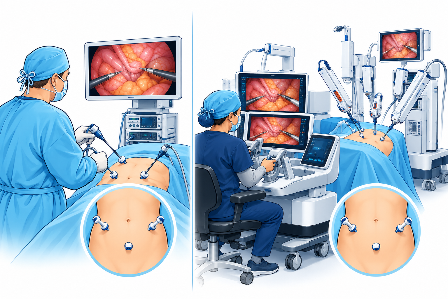
Rectal surgery can be performed via an open approach or a minimally invasive approach depending on the situation.
Laparoscopy allows surgery to be performed using a camera and instruments introduced through small incisions.
Robot-assisted surgery is another minimally invasive approach.
It can notably facilitate certain maneuvers within the narrow space of the pelvis.
The surgeon directly controls the instruments from a console: the robotic system does not perform any maneuver autonomously.
Can immediate surgery sometimes be avoided?
A highly selective option after a complete response to treatment
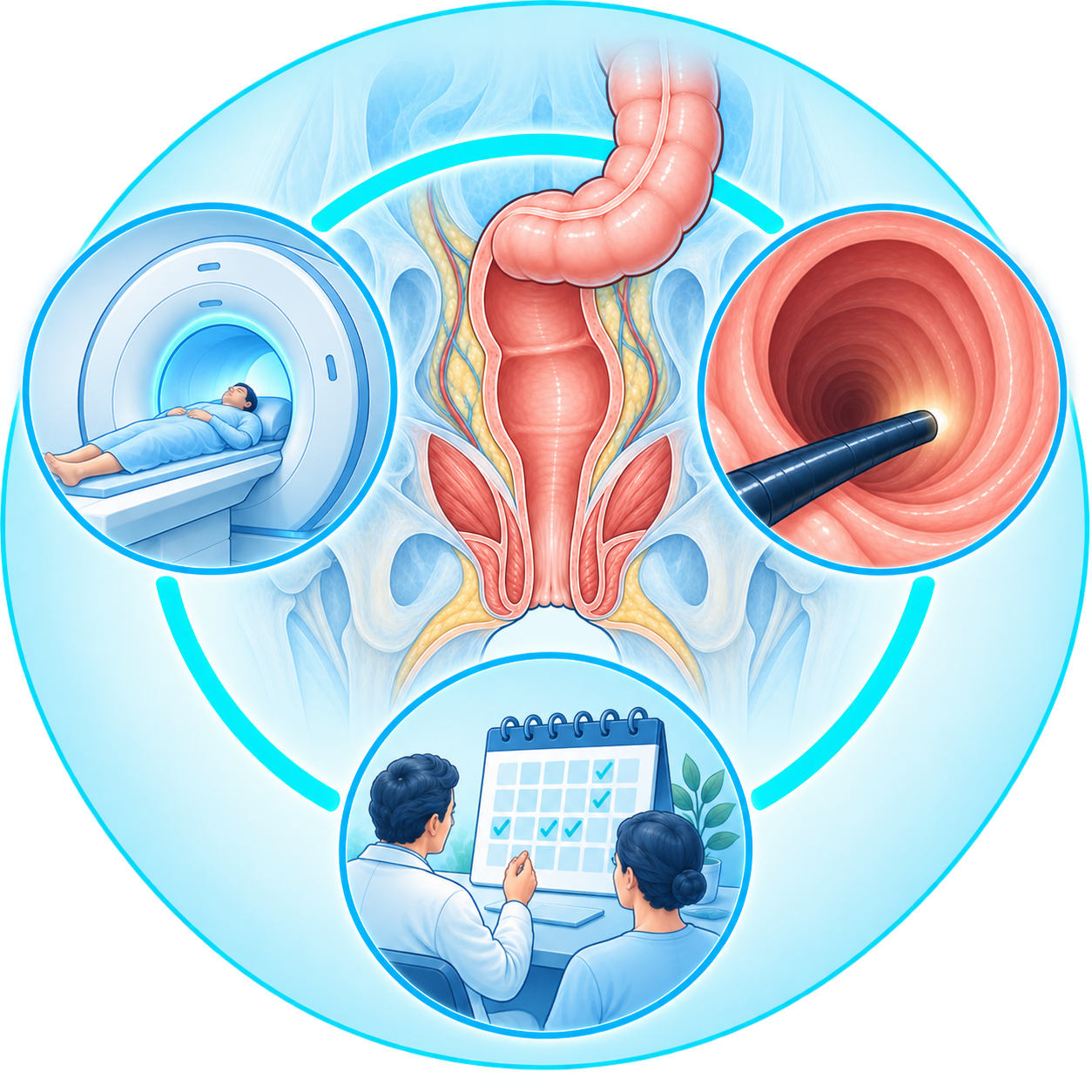
In some patients who have received preoperative treatment, the tumor may show a complete clinical response, meaning no residual tumor is detectable on the exams used to assess response.
In carefully selected situations, a surveillance strategy without immediate surgery may then be discussed.
This approach is often called Watch and Wait.
It does not mean the patient is simply “left without treatment.”
On the contrary, it requires particularly rigorous oncologic surveillance, combining clinical exams, endoscopy, and MRI at regular intervals.
If local regrowth occurs, surgery may be necessary.
This strategy applies only to selected situations and must be discussed within an experienced team.
What happens after surgery?
Pathology analysis clarifies the disease’s characteristics
The surgical specimen is analyzed by the pathologist.
This analysis notably allows study of:
- the residual tumor
- its depth of extension
- the resection margins
- the mesorectum
- the lymph nodes removed
- the response to any preoperative treatment
This information is combined with the data obtained before surgery to determine the next steps in care.
What functional consequences follow rectal surgery?
Bowel function can be altered
After rectal resection with sphincter preservation, bowel function may differ from what it was before surgery.
Some patients may notably experience:
- more frequent bowel movements
- fragmented bowel movements
- more urgent needs
- difficulty delaying evacuation
- a feeling of incomplete emptying
The extent of these changes varies considerably from one patient to another.
It depends notably on the level of the anastomosis, the treatments received, and prior bowel function.
When several of these symptoms occur together, this is sometimes called low anterior resection syndrome, or LARS.
Symptoms can improve gradually, and various measures can be offered when they are bothersome.

A Multidisciplinary Approach
Several specialists contribute to the decision
The management of rectal cancer involves several specialties.
Depending on the situation, the case may notably involve:
- gastroenterologists
- radiologists
- colorectal surgeons
- oncologists
- radiation oncologists
- pathologists
- stoma care nurses when necessary
The goal is to build a strategy tailored to the tumor’s location, its stage, and the patient’s individual characteristics.
A Personalized Surgical Approach
Understanding Precisely the Proposed Procedure
Rectal surgery requires a particularly precise understanding of pelvic anatomy.
The consultation notably makes it possible to explain:
- the exact location of the tumor
- the MRI results
- the possible role of preoperative treatment
- the type of resection considered
- the possibility of preserving the sphincter
- the type of anastomosis
- the possibility of a temporary or permanent stoma
- the proposed surgical approach
- the possible functional consequences
Dr Pierre Fournier
General and Visceral Surgery
[Real photo of Dr Fournier — to be added once available]
Frequently Asked Questions
Both belong to the colorectal cancer family, but the rectum is located within the pelvis. This location affects the local work-up, the treatment strategy, and certain aspects of surgery.
MRI allows precise assessment of the tumor’s local extension, the mesorectum, the lymph nodes, and the relationship with pelvic structures.
No. The need for radiotherapy depends on the tumor’s characteristics and the treatment strategy.
No. Preoperative treatments are tailored to the tumor’s stage and risk profile.
The mesorectum is the fatty tissue surrounding the rectum. It notably contains blood vessels and lymph nodes.
TME, or total mesorectal excision, is an oncologic surgical technique aimed at removing the affected rectum together with its mesorectal envelope in an anatomically precise manner.
No. This depends notably on the tumor’s location, its extension, its relationship with the sphincter, and the ability to achieve a satisfactory oncologic resection.
No. It is not systematic.
A temporary stoma can be used to divert the bowel and protect certain low rectal anastomoses while they heal.
It may notably be necessary when an oncologic resection requires removal of the rectum and anal sphincter.
Yes, in certain situations. The robotic system remains entirely controlled by the surgeon.
This is an intensive surveillance strategy that may be discussed for certain patients with a complete clinical response after preoperative treatment. It requires very rigorous specialized follow-up.
Not necessarily. Rectal resection can alter the frequency, urgency, and fragmentation of bowel movements. The extent of these changes varies by patient and procedure.
Has a rectal cancer just been diagnosed?
The surgical consultation makes it possible to precisely analyze the tumor’s location, the pelvic MRI, the staging work-up, and the proposed treatment strategy. The goal is to understand the different stages of treatment and, when surgery is considered, the type of procedure best suited to the situation.